


Tuberous Sclerosis Complex and Type 1 Neurofibromatosis
The diagnostic test is a Wood light examination. An example of another child with these lesions as they appear under Wood light is shown in Figure B. The Wood light is ultraviolet; it exploits the fact that melanin will preferentially absorb the light and appear darker. Those areas that contain less melanin thus will be highlighted.
Case 1:
This 4-year-old girl was born with these cutaneous lesions. They were overlooked until recently.
What is the underlying disorder--and what clinical diagnostic test would have confirmed the diagnosis at birth?
Case 2:
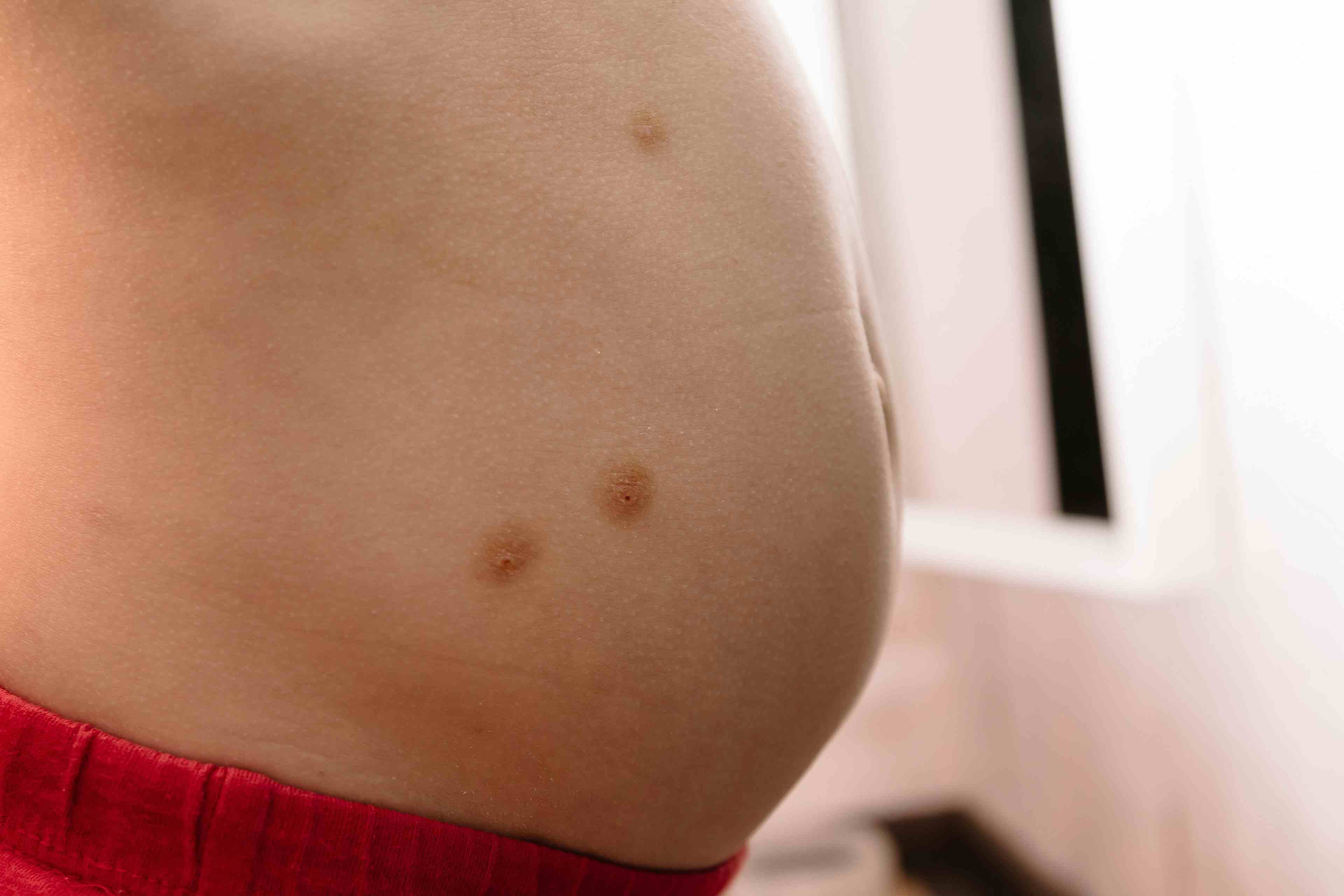
This infant has a number of pigmented macules over her skin.
The absolute number of lesions is important. Why?
Case 1: Tuberous sclerosis complex, confirmed by Wood light examination
The diagnostic test is a Wood light examination. An example of another child with these lesions as they appear under Wood light is shown in Figure B. The Wood light is ultraviolet; it exploits the fact that melanin will preferentially absorb the light and appear darker. Those areas that contain less melanin thus will be highlighted.
The diagnosis is, of course, tuberous sclerosis complex (TSC), and the skin lesions are those of "ash leaf" hypomelanotic macules. These macules are lance-ovate in shape (one end rounded and the other end a sharp point); they measure from 1 to 12 cm in diameter and may number from 1 to 50. They occur predominantly on the lower back and buttocks (A) but are found randomly distributed over the body. They were named by Dr Thomas Fitzpatrick, who thought they resembled the leaves of the eastern mountain ash tree. Nothing is absolute, however. These hypomelanotic macules come in a variety of oval shapes.
It has been shown that 5% of the general population have a single hypomelanotic macule; 1% have 2 lesions, and only 0.1% have 3. Therefore, the 1998 consensus conference on TSC has determined that 3 or more hypomelanotic macules must be present to be considered as a major diagnostic criterion of TSC.1
1.Roach ES, Gomez ER, Northrup H. Tuberous sclerosis complex consensus conference: revised clinical diagnostic criteria. J Child Neurol. 1998;13:624-628.
Case 2: The diagnosis of type 1 neurofibromatosis is based on the number of pigmented macules.
I am commonly asked to review healthy-appearing infants and children who have café-au-lait macules (CALMs) to help determine whether they have neurofibromatosis. The prototypical CALM is a well-circumscribed, evenly pigmented macule that measures from 0.2 to 4 cm at birth and that increases in size proportionate with body growth. In older children, these lesions may be quite large (25 cm or more). They occur predominantly over the lower back and buttocks in infancy. The reported incidence of CALMs in newborns is approximately 2.7%. Macules can be found in 25% to 30% of children.
What do these lesions mean? In infants, I accept 1 macule as normal and more than 3 macules as suspicious for type 1 neurofibromatosis (NF-1). If there are more than 5 macules that are 0.5 cm or larger, I recommend that the child be evaluated with the expectation that he or she does have NF-1. Multiple CALMs are present in more than 90% of children with NF-1. The presence of 6 CALMs that measure 0.5 cm or more in children and more than 1.5 cm in adolescents defines one of the major criteria necessary for the diagnosis of NF-1.
Axillary freckling (Crowe sign) is the presence of multiple 0.5- to 4-mm CALMs in the axilla. These macules appear when a child reaches 3 to 5 years of age and occur in 30% to 50 % of children with NF-1. The presence of both 6 CALMs and axillary freckling allows for the definitive diagnosis of NF-1. *
References:
REFERENCE:


Dupilumab safe, effective for up to 1 year for atopic dermatitis in infants, preschool children
May 3rd 2024According to new study data presented at the 2024 Pediatric Academic Societies Meeting, dupilumab (dupixent; Sanofi and Regeneron) demonstrated positive safety and efficacy results for up to 1 year in infants and preschool-age children with atopic dermatitis.

Recognize & Refer: Hemangiomas in pediatrics
July 17th 2019Contemporary Pediatrics sits down exclusively with Sheila Fallon Friedlander, MD, a professor dermatology and pediatrics, to discuss the one key condition for which she believes community pediatricians should be especially aware-hemangiomas.


Young woman with tick bites presents with erythematous papules, headaches, and fatigue
April 8th 2024A young woman with no significant past medical history returns from hiking with several white-spotted ticks and experiences erythematous papules, rashes, headaches, and fatigue. What’s the diagnosis?