


Neonatal feeding in practice: Nutrition principles for babies with a history of intestinal injury or resection
A history of intestinal injury can create some feeding challenges for infants. Here are some of the causes, feeding guidelines, and approaches to feeding difficulties to use in practice.
Infants who have a history of intestinal injury as neonates can pose challenges for the pediatricians who care for these babies after they are discharged from the hospital. Here are some of the causes, feeding guidelines, and approaches to feeding difficulties that you can use in your practice.
Common causes of neonatal intestinal injury include necrotizing enterocolitis (NEC), spontaneous intestinal perforation, gastroschisis, intestinal atresia, and volvulus (Table 1). Intestinal injury in the neonate typically presents acutely with vomiting, inability to tolerate enteral feeds, abdominal distention, and bloody stools. Treatment typically requires at least the temporary adoption of bowel rest and parenteral nutrition.1


Table 1
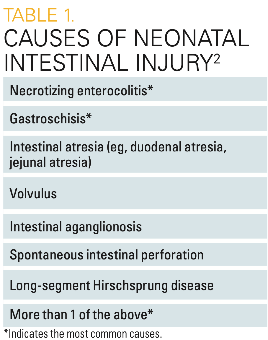
Intestinal failure is generally defined by the loss of normal function of the intestine, regardless of length of residual small bowel, resulting in reliance on parenteral nutrition for calories and fluid to sustain life. NEC and gastroschisis are the most common causes of long-term intestinal failure in infants, which results in significant morbidity, mortality, and cost to families and medical systems.1,2 Those infants who undergo significant intestinal resection and intestinal failure, and thus need long-term parenteral nutrition, require specialized care in multidisciplinary intestinal rehabilitation centers. However, the majority of infants with intestinal injury will tolerate enteral nutrition by hospital discharge.3 Therefore, the majority of these infants will be cared for by their primary pediatricians, who should be aware that these infants are prone to a variety of ongoing nutritional challenges.
Feeding difficulties include poor oral intake, failure to thrive, nutrient deficiencies, gastroesophageal reflux disease (GERD), and intestinal dysmotility. Intestinal dysmotility secondary to resection or injury can cause chronic vomiting, diarrhea, abdominal distention, and small intestinal bacterial overgrowth (SIBO). More than 50% of infants with intestinal injury are prescribed medications to address 1 or more of these issues.2 Overall positive prognostic indicators for successful tolerance of enteral feeds in these infants include the residual length of the small bowel following resection (the longer the residual bowel, the better), the presence of an intact ileocecal valve, and an intact colon; therefore, it is important to understand the infant’s surgical course, residual anatomy, and any neurodevelopmental sequelae.4
General feeding guidelines and principles
The importance of feeding guidelines and algorithms for preterm and other critically ill infants who are at increased risk of NEC are well established.5 However, guidelines for feeding following intestinal injury are not standardized; they can be highly variable across institutions and even among providers at the same institutions.2,6
Enteral feeding stimulates the intestine to heal and grow. Data have shown that early implementation of enteral feeds following intestinal injury (<5-7 days) promotes intestinal adaptation and is not associated with worse outcomes7 or recurrence of NEC. In fact, early feeding with the use of consistent, standardized feeding advancement has been associated with shorter length of stay, quicker achievement of enteral feeding goals, fewer infectious complications, and reduced overall cost to the patient and health system.8,9 For those patients who require surgery for intestinal injury, guidelines support early post-operative enteral feeding with breast milk if available.
Once infants are advanced to full enteral feeds, close monitoring of weight is needed to ensure age-appropriate weight gain. A follow-up study of infants with intestinal injury demonstrated that a majority of these infants will be discharged with some proportion of supplemental tube feeding via nasogastric or gastrostomy tube, but most will be weaned to primarily oral feeds by the time they are aged 1 year.10 Those with a history of NEC and spontaneous intestinal perforation were found to have poor weight gain 3 months after discharge.10 In some studies, poor weight gain in children with surgical NEC persisted through the first 2 years of life.11 Infants with intestinal injury therefore require frequent feeding adjustment and weight checks in those early months to account for higher caloric requirements for optimal growth. Foods that are complementary to formula and breast milk should be added to the diet as developmentally appropriate, with the provision that fruit juices and sugary foods can produce a high solute load in the small bowel. Children with intestinal dysfunction or resection may be more prone to the osmotic diarrhea caused by these foods.
TYPE OF FEEDING
Breast milk has long been favored as the initial choice for feeding infants both with and without intestinal injury or resection. Breast milk reduces the risk of NEC. Breast milk attenuates the proinflammatory pathway that is mediated by a molecular signaling protein, Toll-like receptor-4, which plays a prominent role in the pathogenesis of NEC.12 The exclusive use of preterm formula instead of breast milk might contribute to maturation of less-favorable intestinal flora, and it lacks the protective effects against NEC that are seen in preterm infants who are fed breast milk. If maternal breast milk is not available to infants with intestinal injury, donor breast milk should be used, if available. Donor milk has also been shown to be protective against further intestinal injury, despite the need for pasteurization.7
Breast milk also has a multitude of beneficial bioactive and immunomodulating components that not only provide nutrition but also provide anti-inflammatory and antioxidant compounds.13 Examples include epidermal growth factors, which promote mucosal healing; secretory immunoglobulin A for immune function; lactoferrin to promote enterocyte growth; and prebiotic substrates, such as human milk oligosaccharides (HMOs), which promote a diverse gut flora. HMOs are carbohydrates found in human milk; they have antimicrobial properties that prevent the adhesion of some pathogenic intestinal bacteria. HMOs diminish intestinal leukocyte adhesion and migration, thereby dampening some of the pro- inflammatory processes in the gut. Synthetically produced HMOs are now available, but not all are functionally equivalent to endogenously produced HMOs.14
Despite breast milk’s many benefits, not all infants with intestinal injury will have access to it or will tolerate it. Formula selection should take into consideration the degree of intestinal injury and other comorbidities. A list of infant formulas and their properties is shown in Table 2. Among infants who had significant bowel injury requiring parenteral nutrition, those who were fed amino acid–based formula, also known as elemental formula, or breast milk required parenteral nutrition for a shorter duration, indicating that amino acid–based formula was also well tolerated by such infants.15 The amino acids and medium-chain triglycerides in these formulas are simpler to absorb.


Table 2
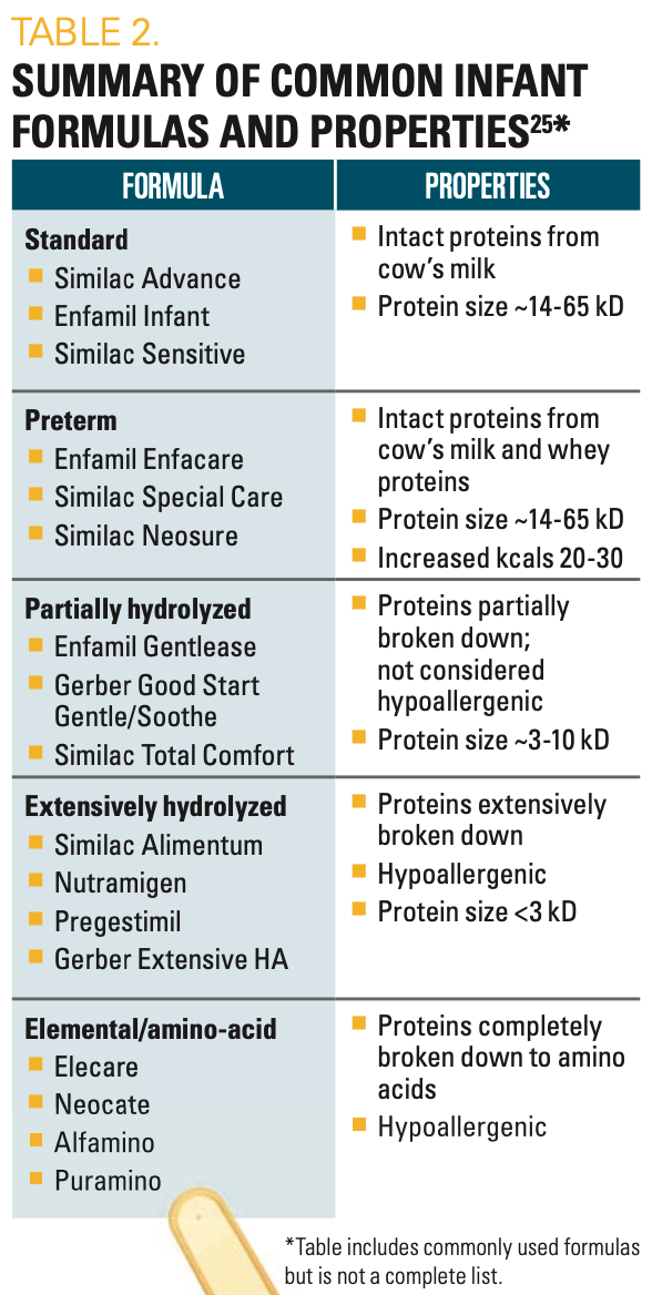
Extensively hydrolyzed formulas should be considered in patients who have suspected milk protein intolerance. Infants who are born extremely prematurely and/or who are critically ill have been found to have worse outcomes with the use of intact-protein, cow’s milk–based formulas.16 These formulas are associated with developing NEC, which can be recurrent; therefore, they should be used cautiously in infants with a NEC history. The elemental formulas (amino-acid based) may be better tolerated; however, more complex intact proteins and other macronutrients promote a greater degree of intestinal growth and adaptation after intestinal resection. Intact cow’s milk proteins may be tolerated later in those infants with more significant bowel injury or resection as the infant’s immune system matures. Term infants with minimal resection may tolerate an intact cow’s milk–based formula initially following intestinal injury.
APPROACH TO FEEDING DIFFICULTIES
Even if they are successfully weaned from parenteral nutrition, infants with bowel injury may still be prone to feeding difficulty due to anatomic changes, dysmotility, and developmental delay. Nearly 60% of infants who experience NEC will have neurodevelopmental delay, which can have a negative impact on their ability to feed orally.1,3 Common symptoms of feeding intolerance include oral aversion, vomiting, fussiness with feeding, abdominal distention, and change in stool frequency and consistency. Treatment and evaluation should be tailored to the infant’s symptoms and medical history.
Certain infants—those with a history of neurodevelopmental delay or who required prolonged periods of bowel rest and parenteral nutrition early in life—may experience poor development of oral-motor coordination or oral aversion. Clinical signs of this include dysphagia, gagging with feeds, or oral feed refusal. Such children might require swallow evaluation with a speech-language pathologist and referral for feeding therapies with a multidisciplinary feeding rehabilitation program.17 Clinical reflux is also a common cause of vomiting and fussiness with feeds. However, the possibilities of delayed gastric emptying or partial obstruction also need consideration, particularly in infants with a history of NEC, gastroschisis, or intestinal reanastomosis; all are prone to dysmotility and strictures.
Radiographic studies can aid in the diagnostic evaluation of anatomic etiologies. They can include (1) abdominal x-ray, to assess for evidence of bowel obstruction or ileus; (2) contrast upper gastrointestinal series or contrast enema to evaluate for stricture, dilatation, or malrotation; and (3) abdominal ultrasound to evaluate both the intestines (intussusception, stricture, or gastric outlet obstruction) and surrounding organs (hydronephrosis).
If no structural cause for vomiting is identified, medications and feeding changes may be indicated. Empiric treatment with 4 to 8 weeks of histamine-2 blockers, proton pump inhibitors (PPIs), or pro-motility agents such as erythromycin can be trialed for suspected reflux. Potential feeding interventions include providing a smaller volume of food with each feeding, more frequent feedings, transitioning to an extensively hydrolyzed or amino acid–based formula, or, ultimately, continuous feeding via nasogastric or gastrostomy tube if other measures fail. Some infants may also require enteral tube feeding due to dysphagia or severe oral aversion. Management of ongoing enteral tube feeding or poor response to formula or medication changes warrants referral to a pediatric dietician and gastroenterologist.
Infants with a history of intestinal injury or bowel resection who experience abdominal distention or bloating without obstruction, feeding intolerance, and changes in stool patterns may have SIBO. Factors that predispose an infant to SIBO include anatomic intestinal abnormalities, dysmotility, bowel resection, ileocecal valve resection, and prolonged use of PPIs that alter the gut flora. In severe cases, SIBO can cause malabsorption, severe diarrhea, acidosis, and altered mental status.
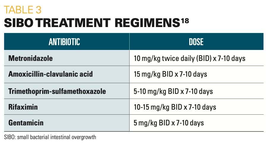
The diagnosis of SIBO can be challenging, as the gold-standard diagnosis technique requires bacterial quantification of jejunal secretions via endoscopy or hydrogen breath testing, neither of which is easily obtained in infants or young children.18 Therefore, empiric treatment with antibiotics is often trialed if there is a strong clinical suspicion of SIBO. A variety of SIBO treatment regimens in children have been proposed (Table 3); however, there is no consensus guideline for pediatric regimens. Empiric treatment is typically prescribed for a 7- to 10-day course, with some infants requiring long-term cycled antibiotics: alternating 1 or 2 antibiotics every 2 to 4 weeks in an effort to prevent development of bacterial resistance. The antibiotic doses for SIBO are typically lower than those in traditional regimens for treating infection, as the goal is to overall reduce the gut bacterial flora.19


Table 3
PROBIOTICS AND PREBIOTICS
There has been great interest in the use of probiotics and prebiotics in infants in recent years. Probiotics are live microorganisms that are administered as an oral supplement to help maintain the balance of beneficial gut bacteria and gut health. Prebiotics are nondigestible oral supplements, usually made up of oligosaccharides, that serve as substrates for nonpathogenic gut bacteria.19 The positive balance of pathogenic and nonpathogenic intestinal microorganisms has an effect on underlying gut inflammation and health.
Preterm infants have later acquisition of and fewer beneficial gut bacteria, including Bifidobacterium and Lactobacillus, when compared with term infants. This lack of diversity is multifactorial and related to mode of delivery, exposure to antibiotics, the hospital environment, and feeding methods.19 The use of probiotics and prebiotics has served to promote and maintain the balance of nonpathogenic gut bacteria, with some data indicating benefits of certain probiotic strains including Lactobacillus rhamnosus, Bacillus infantis, Bacillus lactis, Streptococcus thermophilus, and Bifidobacterium.7,19
Recent American Gastroenterological Association guidelines recommend the use of probiotics in preterm and low-birth-weight infants that contain a combination of Lactobacillus and Bifidobacterium species to prevent mortality and NEC.20 However, the benefit of probiotics in infants following intestinal injury is unclear and, currently, insufficient evidence recommends the routine use of probiotics following intestinal injury.21 There is no consensus regarding the optimal dose or frequency of probiotics or prebiotics in children, and it is important to note that there have been case reports of bacterial translocation and bacteremia associated with probiotic use in infants.22 Although probiotics and prebiotics have shown promising beneficial effects in some pediatric patients, their use should be deter- mined on a case-by-case basis in lower-risk infants.7
NUTRITIONAL DEFICIENCIES AND PITFALLS
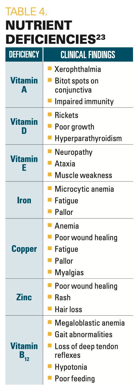
Even those infants with relatively short segments of bowel resection should be monitored for nutrient deficiencies. Risk is related to the particular location of resected bowel and the degree of dysmotility causing diarrhea with malabsorption. For example, copper is absorbed primarily in the proximal small intestine, so resections or dysfunction of the duodenum and proximal jejunum can lead to copper deficiency. Vitamin B12 is absorbed only in the terminal ileum, and B12 deficiency may not become apparent for several months after resection of the terminal ileum. Additional common nutrient deficiencies to be aware of include iron-deficiency anemia, copper-deficiency anemia, and deficiencies of fat-soluble vitamins (A, D, E, K) and of trace minerals such as selenium and zinc (Table 4).23 Depending on clinical signs and symptoms, and on the infant’s pattern of bowel injury, these levels should be monitored 1 to 2 times per year for the first 2 to 3 years, or more frequently if repletion is needed.24


Table 4
Conclusion
Infants with a history of intestinal in- jury from neonatal conditions such as NEC, gastroschisis, and other etiologies of intestinal resection are predisposed to feeding difficulties and nutritional deficiencies. The infant’s surgical history and remaining bowel anatomy need to be considered when monitoring feeding tolerance, growth, and nutritional status. These infants often require the use of hydrolysate or elemental formula if breast milk is not tolerated; however, the use of breast milk should be prioritized given its unique immunologic and probiotic benefits. Feeding intolerance, poor growth, GERD, and dysmotility can also be managed with feeding volume and rate adjustments, and with the assistance of various medications if anatomic etiologies, such as stricture, have been excluded.
References
1. Neu J, Walker WA. Necrotizing enterocolitis. N Engl J Med. 2011;364(3):255-264. doi:10.1056/NEJMra1005408
2. Squires RH, Duggan C, Teitelbaum DH, et al. Pediatric Intestinal Failure Consortium. Natural history of pediatric intestinal failure: initial report from the Pediatric Intestinal Failure Consortium. J Pediatr. 2012;161(4):723-728.e2. doi:10.1016/j.jpeds.2012.03.062
3. Jones IH, Hall NJ. Contemporary outcomes for infants with necrotizing enterocolitis–a systematic review. J Pediatr. 2020;220:86-92.e3. doi:10.1016/j.jpeds.2019.11.011
4. Belza C, Fitzgerald K, de Silva N, et al. Predicting intestinal adaptation in pediatric intestinal failure: a retrospective cohort study. Ann Surg. 2019;269(5):988-993. doi:10.1097/SLA.0000000000002602
5. Wales PW, Allen N, Worthington P, George D, Compher C, American Society for Parenteral and Enteral Nutrition, Teitelbaum D. A.S.P.E.N. clinical guidelines: support of pediatric patients with intestinal failure at risk of parenteral nutrition-associated liver disease. JPEN J Parenter Enteral Nutr. 2014;38(5):538-557. doi:10.1177/0148607114527772
6. Xu JH, Coo H, Fucile S, et al; Canadian Neonatal Network Investigators. A national survey of the enteral feeding practices in Canadian neonatal intensive care units. Paediatr Child Health. 2019;25(8):529-533. doi:10.1093/pch/pxz112
7. Moschino L, Duci M, Fascetti Leon F, et al. Optimizing nutritional strategies to prevent necrotizing enterocolitis and growth failure after bowel resection. Nutrients. 2021;13(2):340. doi:10.3390/nu13020340
8. Shores DR, Mogul D, Allen J, Delarmente BA, Padula W. Cost-effectiveness analysis of feeding guidelines for infants following intestinal surgery. J Pediatr Gastroenterol Nutr. 2020;70(5):657-663. doi:10.1097/MPG.0000000000002642
9. Shakeel F, Newkirk M, Sellers A, Shores DR. Postoperative feeding guidelines improve outcomes in surgical infants. JPEN J Parenter Enteral Nutr. 2020;44(6):1047-1056. doi:10.1002/jpen.1726
10. Varma S, Bartlett EL, Nam L, Shores DR. Use of breast milk and other feeding practices following gastrointestinal surgery in infants. J Pediatr Gastroenterol Nutr. 2019;68(2):264-271. doi:10.1097/MPG.0000000000002128
11. Hintz SR, Kendrick DE, Stoll BJ, et al; NICHD Neonatal Research Network. Neurodevelopmental and growth outcomes of extremely low birth weight infants after necrotizing enterocolitis. Pediatrics. 2005;115(3):696-703. doi:10.1542/peds.2004-0569
12. Good M, Sodhi CP, Egan CE, et al. Breast milk protects against the development of necrotizing enterocolitis through inhibition of Toll-like receptor 4 in the intestinal epithelium via activation of the epidermal growth factor receptor. Mucosal Immunol. 2015;8(5):1166-1179. doi:10.1038/mi.2015.30
13. Hodzic Z, Bolock AM, Good M. The role of mucosal immunity in the pathogenesis of necrotizing enterocolitis. Front Pediatrics. 2017;5:40. doi:10.3389/fped.2017.00040
14. Bode L, Jantscher-Krenn E. Structure-function relationships of human milk oligosaccharides. Adv Nutr. 2012;3(3):383S-391S. doi:10.3945/an.111.001404
15. Andorsky DJ, Lund DP, Lillehei CW, et al. Nutritional and other postoperative management of neonates with short bowel syndrome correlates with clinical outcomes. J Pediatr. 2001;139(1):27-33. doi:10.1067/mpd.2001.114481
16. Abrams SA, Schanler RJ, Lee ML, Rechtman DJ. Greater mortality and morbidity in extremely preterm infants fed a diet containing cow milk protein products. Breastfeed Med. 2014;9(6):281-285. doi:10.1089/bfm.2014.0024
17. Kerzner B, Milano K, MacLean WC Jr, Berall G, Stuart S, Chatoor I. A practical approach to classifying and managing feeding difficulties. Pediatrics. 2015;135(2):344-353. doi:10.1542/peds.2014-1630
18. Malik BA, Xie YY, Wine E, Huynh HQ. Diagnosis and pharmacological management of small intestinal bacterial overgrowth in children with intestinal failure. Can J Gastroenterol. 2011;25(1):41-45. doi:10.1155/2011/604643
19. Mangal Patel R, Wei Denning P. Therapeutic use of prebiotics, probiotics, and postbiotics to prevent necrotizing enterocolitis: what is the current evidence? Clin Perinatol. 2013;40(1):11-25. doi:10.1016/j.clp.2012.12.002
20. Preidis GA, Weizman AV, Kashyap PC, Morgan RL. AGA technical review on the role of probiotics in the management of gastrointestinal disorders. Gastroenterology. 2020;159(2):708-738.e4. doi:10.1053/j.gastro.2020.05.060
21. Fallon EM, Nehra D, Potemkin AK, et al. American Society for Parenteral and Enteral Nutrition (A.S.P.E.N.) Board of Directors, Puder M. A.S.P.E.N. clinical guidelines: nutrition support of neonatal patients at risk for necrotizing enterocolitis. JPEN J Parenter Enteral Nutr. 2012;36(5):506-523. doi:10.1177/0148607112449651
22. Bertelli C, Pillonel T, Torregrossa A, et al. Bifidobacterium longum bacteremia in preterm infants receiving probiotics. Clin Infect Dis. 2015;60(6):924-927. doi:10.1093/cid/ciu946.
23. Diab L, Krebs NF. Vitamin excess and deficiency. Pediatr Rev. 2018;39(4):161-179. doi:10.1542/pir.2016-0068
24. Bielawska B, Allard JP. Parenteral nutrition and intestinal failure. Nutrients. 2017;9(5):466. doi:10.3390/nu9050466
25. Vandenplas Y, Bhatia J, Shamir R, et al. Hydrolysed formulas for allergy prevention. J Pediatr Gastroenterol Nutrition. 2014;58(5):549-552. doi:10.1097/MPG.0000000000000318


The benefits of concurrent fetal and maternal heart rate monitoring
April 17th 2024A recent study revealed that employing maternal heart rate monitoring alongside fetal heart rate monitoring during labor significantly decreases the incidence of neonatal encephalopathy and severe neonatal acidemia.