


Lymphangioma Circumscriptum With Secondary Cellulitis
For 3 days, a 6-year-old boy had “redness and pain” of skin on his left upper abdomen. Physical examination revealed a large cluster of vesicles with underlying erythema and mild warmth.

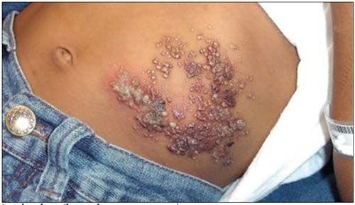

For 3 days, a 6-year-old boy had “redness and pain” of skin on his left upper abdomen. Physical examination revealed a large cluster of vesicles with underlying erythema and mild warmth. The patient’s mother stated that the lesion had been present for 5 years, that it had been diagnosed as lymphangioma circumscriptum by a dermatologist, and that excision was pending. Secondary cellulitis of the existing lesion was considered, and an oral antibiotic was prescribed. At follow-up, signs of infection had cleared.
Lymphangiomas, which make up about 4% of all vascular tumors and 26% of benign vascular tumors in children, are divided into 4 main types: lymphangioma circumscriptum, cavernous lymphangioma, cystic hygroma, and benign lymphangioendothelioma.1 Lymphangioma circumscriptum is a rare type caused by dilation of subcutaneous lymphatic cisterns, which leads to outpouchings that protrude through the skin and form vesicles.2 Half of cases present at birth, 90% by age 2 years.3
Clinically, the disorder can be described as clusters of small (usually 1- to 2-mm) translucent vesicles; hemorrhage in and around the vesicles may cause the lesions to appear pink, red, or black.2 The lesions can occur anywhere on the skin or mucous membranes, but they most commonly develop on the proximal extremities, trunk, axillae, and buttocks.1 The differential diagnosis can include warts, molluscum contagiosum, hemangioma, angiokeratoma, lymphangioendothelioma, lymphangiectasis, metastatic carcinoma of the skin, herpes simplex, herpes zoster, dermatitis herpetiformis, and malignant melanoma.2,3
Treatment may be considered for cosmesis, pain, swelling, and recurrent cellulitic infections.3 Options include cryotherapy, laser therapy, sclerosing agents, and suction-assisted lipectomy; however, surgical resection using MRI to obtain adequate margins is the preferred method.2
References:
REFERENCES:
1. Heller M, Mengden S. Lymphangioma circumscriptum. Dermatol Online J. 2008;14:27.
2. Mordehai J, Kurzbart E, Shinhar D, et al. Lymphangioma circumscriptum. Pediatr Surg Int. 1998;13:208-210.
3. Patel GA, Schwartz RA. Cutaneous lymphangioma circumscriptum: frog spawn on the skin. Int J Dermatol. 2009;48:1290-1295.


Recognize & Refer: Hemangiomas in pediatrics
July 17th 2019Contemporary Pediatrics sits down exclusively with Sheila Fallon Friedlander, MD, a professor dermatology and pediatrics, to discuss the one key condition for which she believes community pediatricians should be especially aware-hemangiomas.


Young woman with tick bites presents with erythematous papules, headaches, and fatigue
April 8th 2024A young woman with no significant past medical history returns from hiking with several white-spotted ticks and experiences erythematous papules, rashes, headaches, and fatigue. What’s the diagnosis?

Tapinarof cream 1% demonstrates efficacy in atopic dermatitis patients with skin of color
March 8th 2024Data from a pair of identical, phase 3, double-blind, randomized, and vehicle-controlled trials were presented at the 2024 American Academy of Dermatology (AAD) Annual Meeting in San Diego, California.