


Incontinentia Pigmenti and Hypomelanosis of Ito
This skin condition is present at birth and usually has significant systemic manifestations. Would you be able to identify it in the nursery?
Case 1:
This skin condition is present at birth and usually has significant systemic manifestations. Would you be able to identify it in the nursery?
Case 2:
These color changes have been present on this child's skin since her birth. Her parents wonder whether anything can be done to "even out" the coloring.
What is this condition--and what will you tell the child's parents?
Case 1: Incontinentia pigmenti
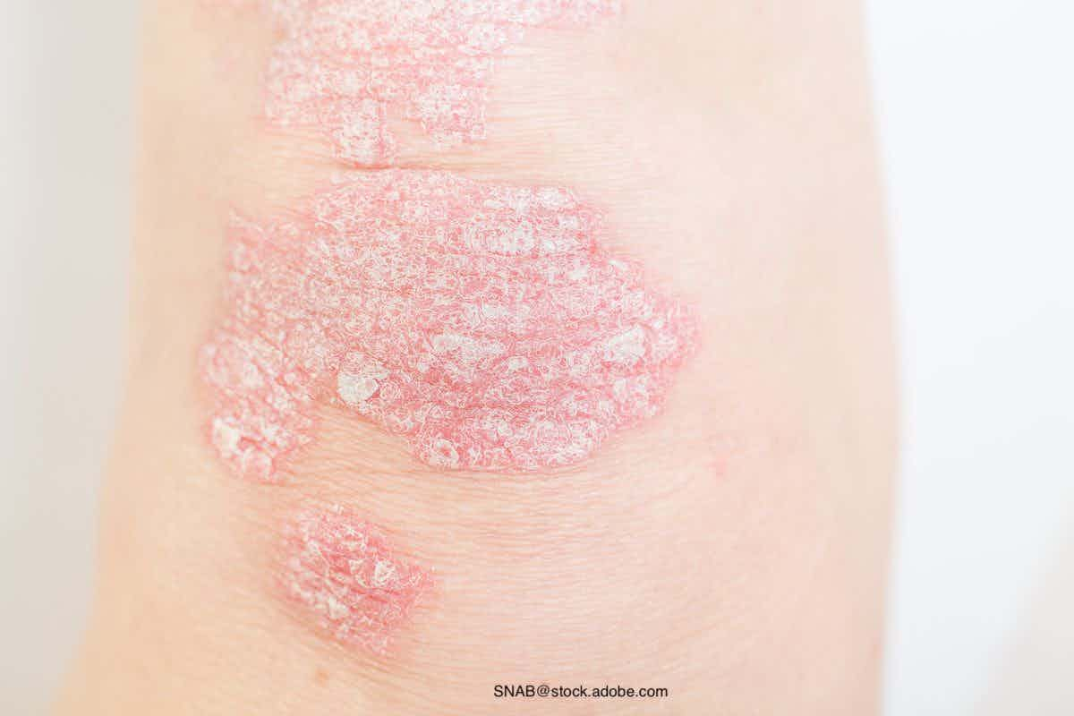
Incontinentia pigmenti is an X-linked dominant disorder that affects the skin, bones, and nerves. The manifestations are present at birth, and the cutaneous findings are characteristic.
The lesions have 4 distinct phases that may overlap or manifest sequentially as follows:
1. Erythema, vesicles, pustules.
2. Verrucous and hyperkeratotic papules.
3. Hyperpigmentation.
4. Hypopigmentation, atrophy, scarring.
The first (inflammatory) phase is usually present at or within 2 weeks of birth. Lesions must be differentiated from other blistering diseases of the newborn period--especially infections, such as herpes simplex. The blisters appear on an erythematous base and develop in crops over the trunk and extremities, often along the lines of Blaschko. A biopsy specimen taken during this phase will show epidermal vesicles with eosinophils as the predominant type of inflammatory cell. This phase generally resolves within the first 6 months of life.
The second (keratotic) phase is characterized by warty papules that appear in a linear pattern over the extremities. These papules may overlap with the inflammatory phase and generally resolve spontaneously within the first 2 years of life (A, B).
The third (hyperpigmented) phase is the most familiar. Linear bands of brown, blue, or gray hyperpigmentation appear in whorls and swirls that conform to the lines of Blaschko. The pigmentation usually continues to appear over the first 2 years of life: it then stabilizes and begins to fade as puberty approaches. Most children lose their pigmentation entirely. The pigmentation does not necessarily follow the exact pattern of the first 2 phases, but will follow the lines of Blaschko.
Atrophy, scarring, and hypopigmentation are the predominant clinical features during the fourth phase. Lesions can occur anywhere on the body.
Dental (75%), neurological (30%), and ophthalmological (35%) manifestations are the most commonly associated systemic issues. Children whose condition is diagnosed in the nursery should be assessed by an ophthalmologist. Retinal vascular proliferation is the most common ocular finding and may result in retinal detachment and blindness.
Case 2: Hypomelanosis of Ito
This child has hypomelanosis of Ito, a manifestation of pigmentary mosaicism. The hypopigmentation, the only cutaneous abnormality, is present at birth. The morphology is described as "splashed paint," and large surface areas are usually involved. Affected children do not typically have any associated systemic abnormalities, but there are rare reports of asso-ciated ocular and neurological abnormalities.
The pigmentary changes follow the lines of Blaschko. These lines were described in 1901 by Dr Alfred Blaschko when he presented his observations on linear dermatosis. He noted that the lines did not follow any known anatomic structures. Subsequent studies appear to prove his hypothesis that these lines have an embryonic origin.1
The lines of Blaschko form a "V" over the vertebral column, an open "S" shape on the sides of the torso and lower abdomen, and an inverted "U" on the anterior arm and adjacent chest wall. They assume a linear (nondermatomal) pattern on the extremities.
There is currently no way to alter the pigmentation. *
References:
REFERENCE:
1.
Happle R. Mosaicism in human skin. Understanding the patterns and mechanisms.
Arch Dermatol.
1993; 129:1460-1470.


Dupilumab safe, effective for up to 1 year for atopic dermatitis in infants, preschool children
May 3rd 2024According to new study data presented at the 2024 Pediatric Academic Societies Meeting, dupilumab (dupixent; Sanofi and Regeneron) demonstrated positive safety and efficacy results for up to 1 year in infants and preschool-age children with atopic dermatitis.

Recognize & Refer: Hemangiomas in pediatrics
July 17th 2019Contemporary Pediatrics sits down exclusively with Sheila Fallon Friedlander, MD, a professor dermatology and pediatrics, to discuss the one key condition for which she believes community pediatricians should be especially aware-hemangiomas.


Young woman with tick bites presents with erythematous papules, headaches, and fatigue
April 8th 2024A young woman with no significant past medical history returns from hiking with several white-spotted ticks and experiences erythematous papules, rashes, headaches, and fatigue. What’s the diagnosis?