


- Pharmacology
- Allergy, Immunology, and ENT
- Cardiology
- Emergency Medicine
- Endocrinology
- Adolescent Medicine
- Gastroenterology
- Infectious Diseases
- Neurology
- OB/GYN
- Practice Improvement
- Gynecology
- Respiratory
- Dermatology
- Mental, Behavioral and Development Health
- Oncology
- Rheumatology
- Sexual Health
- Pain
Genital Lesions: A Photo Essay
Phimosis, paraphimosis, hypospadias, and congenital hemagioma are just a few of the congenital anomalies that affect the genitals of newborns and young children.
The most common congenital anomaly of the penis, hypospadias is characterized by a urethral meatus that is ectopically located proximal to the normal location and on the ventral aspect of the penis.


Courtesy of Alexander K.C. Leung and C Pion Kao, MD.
In this case of phimosis in a 5-year old boy, the preputial opening of the penis is minute and the foreskin non-retractable. Phimosis is physiological at birth and may result from narrowing or scarring of the preputial opening. Ballooning of the foreskin when the child voids-- balanoposthitis –is a key clinical sign


Courtesy of Alexander K.C. Leung and C Pion Kao, MD.
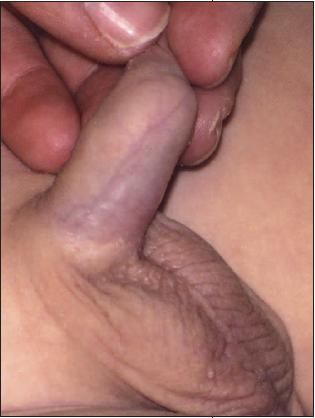
Paraphimosis develops when a phimotic foreskin (see previous image) is retracted past the coronal sulcus and becomes trapped. Lymphatic and venous stasis develops, with resultant pain and edema of the foreskin. The constricting band proximal to the swelling is pathognomonic.


Hidradenitis suppurativa, also referred to as acne inversa or Verneuil disease, involves a chronic, suppurative inflammation of the apocrine glands in the perineum, axilla, and submammary and perianal regions. Initial occlusion of hair follicles allows sweat to accumulate beneath keratin plugs which promotes bacterial proliferation. The most common causative microorganism is Staphylococcus aureus. Women are more often affected than men. This 17-year-old was obese.


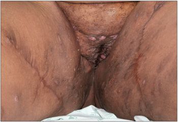
Courtesy of Himalee Sabins, MD, and Deepak Kamat, MD, PhD
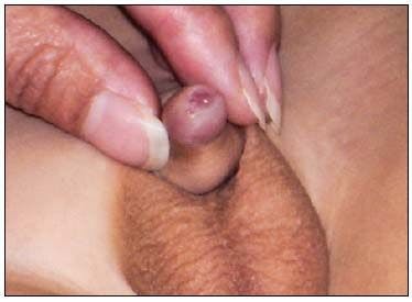
The whitish mass seen here under the foreskin of a 5-year-old boy is an accumulation of desquamated epithelial cells called smegma. Lesions are characteristically smooth, round, uniform, doughy, well-circumscribed, and located in the midshaft of the penis. Smegma helps to dissect the space between the glans and foreskin and also prevents readherence.


Courtesy of Alexander K.C. Leung and C Pion Kao, MD.
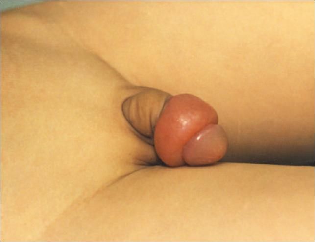
This genital mass in a 6-month old led to concerns of sexual abuse. The mass was circumscribed, compressible, and warm. It was diagnosed as an infantile genital hemangioma, deep variety. Such lesions can be diagnostically challenging and may be mistaken for child maltreatment because of their varied appearance and presentation.


Courtesy of Susan Bradford, MD.
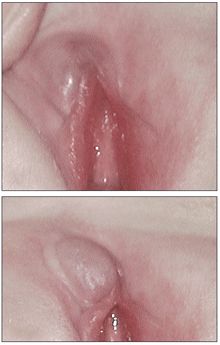
Parents of a 4-year-old boy noted this tiny nontender cystic mass (diameter, 2 mm) on the glans near the urethral meatus. Following excision, histologic examination showed that the cyst was lined by columnar epithelium. The cause of such parameatal urethral cysts is unclear. Congenital lesions however, are believed to be caused by faulty separation of the foreskin from the glans or occlusion of the parameatal duct.


Courtesy of Alexander K.C. Leung and C Pion Kao, MD.